Home / Health / 3D Imaging Revolutionizes Heart Surgery, Saving Lives
3D Imaging Revolutionizes Heart Surgery, Saving Lives
12 Nov
Summary
- 3D imaging helps surgeons plan and perform life-saving heart operations
- 3D models allow doctors to visualize heart and blood vessels from all angles
- 3D-printed artery models help practice procedures before surgery
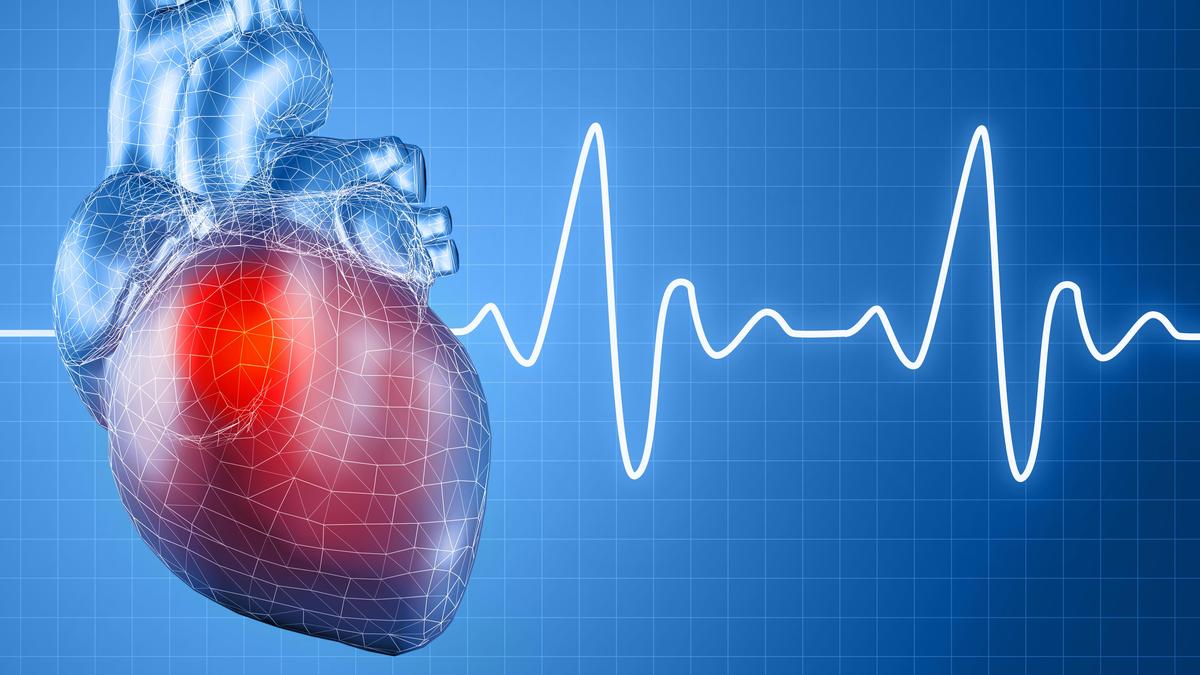
As of November 2025, 3D imaging has become a crucial tool in modern heart surgery, helping doctors save lives with unprecedented precision. By combining data from CT and MRI scans, surgeons can now create lifelike, digital 3D models of the heart and its surrounding blood vessels. Unlike traditional 2D images, these 3D renderings can be rotated, enlarged, and studied from every angle, allowing doctors to thoroughly understand a patient's unique anatomy even before the operation begins.
This technology has proven especially valuable in treating conditions like aortic dissection and aneurysm, where every millimeter matters. Surgeons can use the 3D models to precisely map the size, location, and extent of the problem, enabling them to plan the optimal surgical approach. In some cases, hospitals even create 3D-printed replicas of the patient's arteries, allowing the team to practice the procedure on a realistic model before the actual operation.
Beyond planning, 3D imaging is also making its way into the operating room itself. Surgeons can now view live, reconstructed 3D images while performing procedures, ensuring devices are placed exactly where they should be. This real-time guidance adds an extra layer of safety and confidence, leading to better outcomes for patients. As this cutting-edge technology continues to evolve, the future of heart surgery is clearly moving toward ever-greater visualization and precision, transforming what was once guided by intuition into a science-driven, personalized approach to healing.